
Bowleg Quick Links: Overview | What is Bowleg | Bowleg Symptoms | Causes of Bowlegs | Blount’s Disease | Diagnosing Bowlegs | Treatment

Bowleg Treatment Videos
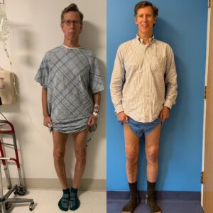
Josh at age 18 chose to have his #bowlegs #bowleg #bowlegscorrection #bowlegsurgery to improve pain, gait, aesthetics, biomechanics, and to prevent knee arthritis in the future. After bilateral surgery, he is walking unassisted at 6 weeks postop. Great to have Dr. Taylor Stauffer @hssresidency rotating on our service #Rozbruch #aaos #llrs #orthopedicsurgery #liveyourbestlife @all4ortho #orthopedicsurgeon #hospitalforspecialsurgery #limblengthening @hspecialsurgery @limbdeformity @reifmd #SRobertRozbruchMD www.limblengthening.com www.hss.edu/limblengthening www.hss.edu/LSARC #hssLLCRS #HSSlimbLengthening

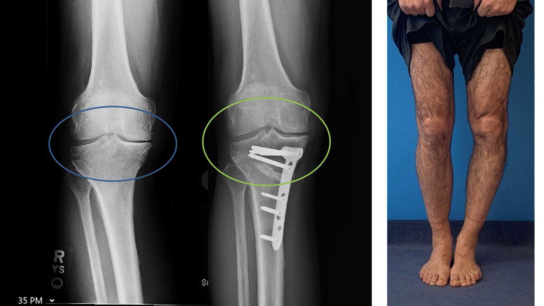
This is my most common technique for osteotomy to correct moderate #bowlegs #bowleg #bowlegged #bowlegscorrection #Rozbruch #aaos #llrs #orthopedicsurgery #liveyourbestlife @all4ortho #orthopedicsurgeon #hospitalforspecialsurgery #limblengthening @hspecialsurgery @limbdeformity @reifmd #SRobertRozbruchMD www.limblengthening.com www.hss.edu/limblengthening www.hss.edu/LSARC #hssLLCRS #HSSlimbLengthening
Michael with severe #blountsdisease 48 degrees #bowleg treated with both intraarticular osteotomy to elevate plateau and metaphasyl osteotomy and gradual correction with a hexapod frame. Stay tuned for second side and outcomes. #Rozbruch #aaos #llrs #orthopedicsurgery #liveyourbestlife @all4ortho #orthopedicsurgeon #hospitalforspecialsurgery #limblengthening @hspecialsurgery @limbdeformity @reifmd #SRobertRozbruchMD www.limblengthening.com www.hss.edu/limblengthening www.hss.edu/LSARC #hssLLCRS #HSSlimbLengthening
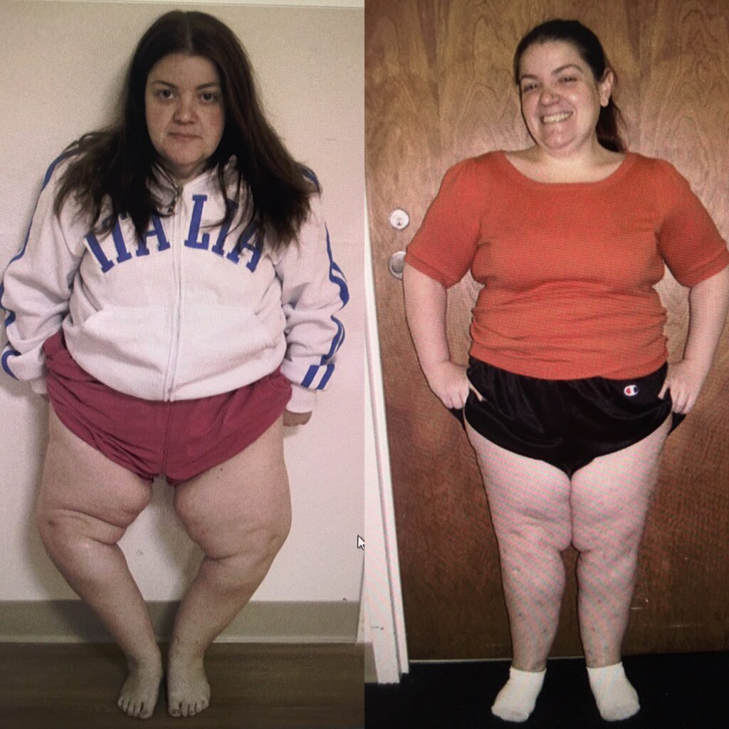
Manuel from #dominicanrepublic with large LEFT #bowleg This was treated with 2 level #bowlegscorrection #bowlegsurgery and 5 months Postop he is very happy. Walks better and knee pain is minimal. #Rozbruch #aaos #llrs #orthopedicsurgery #liveyourbestlife @all4ortho #orthopedicsurgeon #hospitalforspecialsurgery #limblengthening @hspecialsurgery @limbdeformity @reifmd #SRobertRozbruchMD www.limblengthening.com www.hss.edu/limblengthening www.hss.edu/LSARC #hssLLCRS #HSSlimbLengthening
Multiple ways you could tackle this developmental biplanar bowleg deformity. It’s a lot to attempt through one osteotomy given the gentle curve of deformity throughout the bone. I chose to correct the shaft/diaphyseal deformity first with long nails, then remove those and dial in the final correction with a medial opening wedge osteotomy and plate. She just came off crutches and looks great! Plates will be removed too of course.
#hssllcrs #hsslimblengthening #bowlegs #bone #bonedeformity #orthopedicsurgery #deformity #orthopedicsurgeon #femur #femurbone #deformitycorrection @bodycad_
A recent publication from #hssllcrs describing our safety performing #tibia #osteotomy . Techniques and outcomes are summarized. This was published in a @aaos_1 network journal. Coauthors include @limblengthening @limbdeformity @reifmd @adamgeffner
.
.
.
#all4ortho #hspecialsurgery #hsslimblengthening #bowleg #knockknees
For this tibial #deformity I utilized #robotic hexapod struts to gradually correct the deformity, so the patient did not have to manually adjust the #externalfixation during treatment. They were so thrilled with the result a lovely clock was gifted to my office!
#hssllcrs #hsslimblengthening #limbdeformity #hexapod #hexapodrobot #bonedeformity #tibiavara #orthopedics #orthopedicsurgery #bowleg #bowlegsurgery @depuysynthestrauma
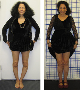
Initially skeptical about surgical correction for knock knees, I eventually convinced this patient she would love her new legs, and she does! She walks with significantly more confidence and wishes she’d done this sooner!
#hssllcrs #hsslimblengthening #bowlegs #knockknees #deformity #orthopedics #orthopedicsurgery @bodycad_ #bonedeformity #surgery #patientspecificplanning
Bowleg Overview
 Bowleg or genu varum is a mal-alignment around the knee that can affect people of all ages, has various causes, and leads to pain and degeneration of the knee if left untreated. The causes can be congenital, developmental, or post-traumatic. The common theme that pervades all age groups is that the knee is abnormally loaded which can lead to pain, increasing deformity, instability, and progressive degeneration. Correction of the deformity leads to improved knee mechanics, better walking, less pain, and prevents the rapid progression of damage to the knee.
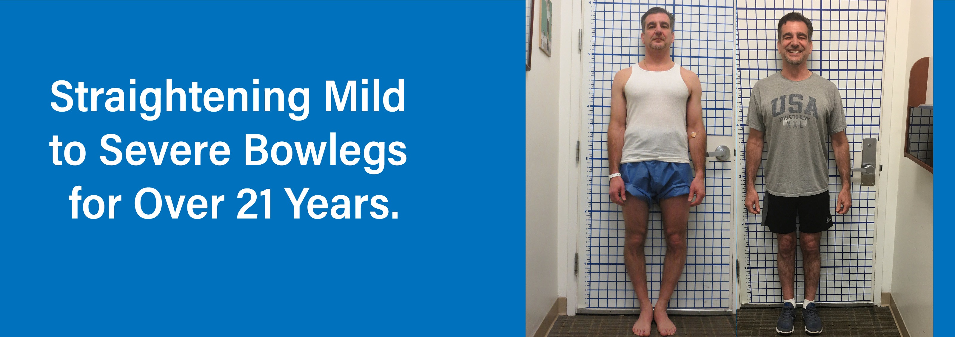
Adult patients who have had bowleg for many years overload the inside (medial compartment) and stretch the outside (lateral collateral ligament) leading to pain, instability, and arthritis. To prevent and delay the need for joint replacement, knee realignment should be done with osteotomy.
Bowleg syndrome causes a person’s legs to appear bowed, bending outward even when the ankles are together. While bowlegs are common in infants, who are often born bowlegged due to their folded position in the womb, in typical growth patterns the child will outgrow this as they start to stand and walk (12 to 18 months old). If a child still has bowlegs at age three or beyond, evaluation for potential deformities may be needed.
What is bowleg?
Bowleg is an orthopedic condition affecting the legs in which a person’s knees stay widely apart even when their ankles are together, giving the legs a bowed look. In many cases, this is due to abnormal bone development, illness, or growth plate deformities.
Up until the age of two, bowing of the legs is not unusual. In fact, there is a broad spectrum of what is considered normal. Doctors frequently use the generic term physiologic genu varum to describe this condition. In common usage, it is also sometimes referred to as bowed legs or bowleggedness.
Infants are typically born bowlegged due to their positioning while in the mother’s womb. In most cases, the legs begin to straighten once the child starts to bear weight on their legs while standing or walking (between 12 and 18 months old). By age two or three, the legs typically begin to look more like knock knees, or knees bending inward toward each other. After six years of age, most children’s knees assume a straighter alignment that is considered normal.
A doctor should be consulted to determine whether a child has the potential to develop a deformity if:
- The angle of the thigh bone to shin bone fall out is outside of the normal range
- The direction of the knees versus the direction the foot falls outside the normal pattern
- Leg is significantly more (or less) angled than the other
What are the symptoms of bowleg?
Symptoms of bowleg syndrome include:
- Bowing of the legs that do not improve after the age of three
- Knees do not touch when standing with feet and ankles together
- Knee or hip pain
- Reduced range of motion in hips
- Difficulty walking or running
- Knee instability
- Progressive knee arthritis in adults
- Patients or parents may be unhappy with aesthetics
What causes bowleg?
Bowleg syndrome may be caused by various illnesses, including:
- Abnormal bone development (bone dysplasia)
- Blount’s disease (more information below)
- Paget’s disease (a metabolic disease impacting the way bones break down and rebuild)
- Improperly healed fractures
- Lead poisoning
- Fluoride poisoning
- Achondroplasia (the most common form of dwarfism)
- Rickets (a bone-weakening disorder caused by a vitamin D deficiency)
- Damage to growth plate
Blount’s disease
Blount’s disease (also called tibia vara) is a growth disorder that affects the growth plates in the bones near the inside of the knee. Blount’s disease causes the growth plate to slow down or stop producing new bone, even as the growth plate near the outside of the knee continues to grow normally. This produces a bowlegged appearance and can cause pain or instability of the knee.
The deformity occurs when the lateral (outer) side of the tibia continues to grow while the medial (inner) side of the bone does not. Blount’s disease may affect one or both legs. Most commonly, the growth deformity is found at the top of the tibia, which is the larger of the two bones in the lower leg. Blount’s disease may affect one or both legs.
Blount’s may be diagnosed in children aged two and up. Depending on the age at diagnosis, a child may have the infantile or adolescent type of this disease. Those with the infantile form of the disease are often early walkers. Obesity is frequently seen in children who have Blount’s, irrespective of the patient’s age. Adults who have not had treatment or inadequate treatment as children can present with large deformity and knee pain and degeneration.
How is Bowleg Mal-alignment diagnosed?
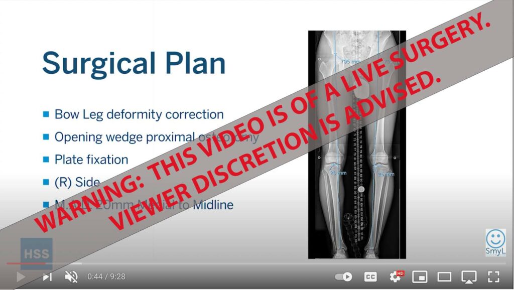
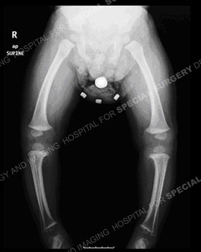
Diagnosis typically includes a history and physical examination as well as a standing alignment X-ray. A standing alignment X-ray or EOS image produces an image of the leg from hip to ankle which helps the orthopedist locate the mechanical axis of the deformity as well as its location. On the x-rays, the magnitude and location of the deformity is identified.
How is bowleg treated?
If it is determined that a child has bowleg syndrome, medical treatment may be necessary. If the deformity is mild, it may be first carefully observed overtime through pediatric orthopedic exams.
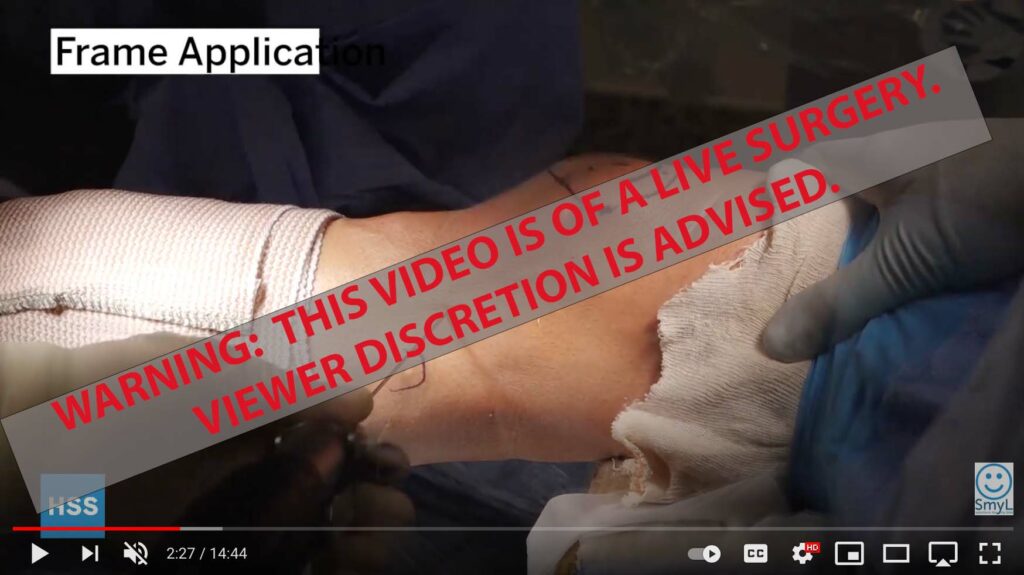
 In some cases, bracing is used as an initial treatment. If a gradual correction does not occur, surgery may be recommended. In the growing child, guided growth minimal incision surgery may be used to encourage the limb to gradually grow straight. Osteotomy, in which the tibia and fibula are each cut, and then realigned may be needed. In some cases, the surgeon will also place an external fixator. With external fixators, pins are inserted into the bone and protrude out of the body to attach to an external stabilizing structure.
Physical therapy is also an important part of treatment, especially in cases where surgery has occurred.
In skeletally mature adolescents and in adults, osteotomy is the treatment to straighten the leg. X-rays are used to determine the location and magnitude of the deformity. In most cases, the tibia is treated, but there are situations when the femur or both femur and tibia are treated. When there is moderate deformity, the osteotomy is typically stabilized with internal fixation (plate or rod). When the mal-alignment is more severe, gradual realignment of the limb through the osteotomy is done with an external fixator.
Get more information about bowleg syndrome, identifying bowleg conditions, treatments, and patient stories:
- Ask the Expert: Why Should I Correct my Bowlegs
- Patient Stories
- Bowleg Case Histories
- Dr. Rozbruch Discusses His Approach to Bowleg Correction